Chapter 11
Skeletal System
- Overview of skeletal system
- Bone growth, remodeling, and repair
3. Bones of the axial skeleton
4. Bones of the Appendicular Skeleton
- Articulations
- Picture References
- Overview of skeletal system
- The human body has 206 bones
- The skeletal system consists of the bones along with the cartilage and fibrous connective tissue, which occurs in the ligament at the joints.
- Function of the Skeleton
- The skeleton supports the body
- Protects soft body parts
- Produces blood cells
- Stores mineral and fat
- Along with the muscles, permits flexible body movement
- Anatomy of a Long Bone


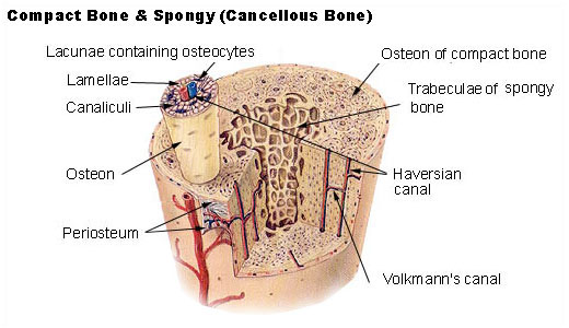
Picture of A Long Bone Picture of the compact bone and Spongy
- The main portion of the bone is called the diaphysis.
- Compact bone is highly organized and composed of tubular units called osteons.
- Spongy bone has an unorganized appearance. It contains numerous thin plates separated by unequal spaces.
- Cartilage is not as strong as bone, but it is more flexible because the matrix gel-like and contains many collagenous and elastic fibers.
o Cartilage has no nerves and no blood vessels, making it harder to heal.
o Three types of cartilage:
1. Hyaline cartilage covers the end of the long bone. Is firm and somewhat flexible.
2. Fibro cartilage is stronger than hyaline cartilage because the matrix contains wide rows of thick, collagen fibers.
3. Elastic cartilage is more flexible than hyaline cartilage because the matrix contains mostly elastin fibers.
- Fibrous connective tissue contains rows of cells called fibroblast separated by bundle of collagenous fibers. This tissue makes up ligaments that connect bone to bone and tendons that connect muscles to a bone at joints.

- Bones can grow throughout a lifetime because they are able to respond to stress by changing size, shape, and strength.
- Bones are composed of living tissues
- Cells involved in growth, remodeling, and repair of the bone are:
o Osteblasts-are bone-forming cells. They secrete the organic matrix of bone and promote the deposition of calcium salts into the matrix.
o Osteocytes-are mature bone cells derived from osteoblasts. They maintain structure of bone.
o Osteoclasts-are bone-absorbing cells. They break down bone and assist in depositing calcium and phosphate in the blood.
- Bone Development and Growth
- Ossification refers to the formation of bone.
- Intramembranous ossification: bones develop between sheets of fibrous connective tissue.
- Endochondral ossification: cartilaginous models of the bones are replaced by calcified bone matrix.
- Bone growth is affected by vitamin D, growth hormone, and sex hormones.
- Bone Remodeling and Its Role in Homeostasis
- Bone remodeling is the renewal of bone. Osteoclasts break down bone and osteoblasts re-form bone. Some bone is recycled each year.
- Bone recycling allows the body to regulate blood calcium.
- Bone remodeling also accounts for why bones can respond to stress.
- Bone Repair
- Repair of a fracture required four steps:
o Hematoma formation
o Fibrocartilginous callus
o Bony callus

The Human Skeleton
- Bones of the axial skeleton
- The axial skeleton consists of:
o The skull
o The hyoid bone
o The vertebral column,


The Human Skull Bones of the Face


The Vertebral Column The Rib Cage
- The skull is formed by the cranium, which protects the brain and the facial bones.
- The hyoid bone anchors the tongue and is the site of attachment of muscles involved with swallowing.
- The vertebral column is composed of vertebrae separated by shock-absorbing disks, which make the column flexible. It supports the head and truck, protects the spinal cord, and is a site for muscle attachment.
- The vertebral column consists of 33 vertebrae.
- Rib Cage
- The rib cage is composed of the thoracic vertebrae, ribs, costal cartilages, and sternum. It protects the heart and lungs.
- The rib cage is part of the axial skeleton.
- Protects the heart and lungs
- Bones of the Appendicular Skeleton
- The appendicular skeleton consists of the bones within the pectoral and pelvis girdles and their attached limbs.
- The bodies have left and right pectoral girdles; each consists of a scapula and clavicle.
- The pectoral girdles and upper limbs are adapted for flexibility.
- The bones in the pectoral girdle are:
o Scapula, clavicle
o The upper limb: humerus, radius, ulna, carpals, metacarpals, phalanges
- The pelvic girdle and the lower limbs are adapted for supporting weight; the femur is the longest and strongest bone in the body.
- The bones in the pelvis girdle are:
o Two coxal bones
o The lower limb: Femur, patella, tibia, fibula, tarsals, metatarsals, and phalanges.
- Articulations
- Bones are joined at joints, of which are thee types:
o Fibrous joints-are immovable
o Cartilaginous joints- is slightly movable.
o Synovial joints – are freely movable.
- Movements Permitted by Synovial Joints
- Intact skeletal muscles are attached to bones by tendons that span joints. When a muscle contracts, one bone moves in relation to another bone.
http://academic.kellogg.cc.mi.us/herbrandsonc/bio201_McKinley/f6-4c_gross_anatomy_of__c.jpg
http://www.fluoridealert.org/pesticides/compact.spongy.bone.jpg?4abcc000
http://classes.midlandstech.com/bio112/figure7.5endochondral%20ossification.jpg
http://library.thinkquest.org/J0111100/graphics/skull1.JPG
http://biology.clc.uc.edu/graphics/bio105/skeleton.jpg
http://services.epnet.com/GetImage.aspx/getImage.aspx?ImageIID=4826
http://academic.kellogg.cc.mi.us/herbrandsonc/bio201_McKinley/f7-28_vertebral_column_c.jpg
http://www.medicalook.com/systems_images/Rib_cage.jpg
Chapter 12
Muscular System
- Overview of Muscular System
- Skeletal Muscle Fiber Contraction
- Whole Muscle Contraction
- Muscular Disorders
- Homeostasis
- Picture References
- Overview of Muscular System
- Type Of Muscles

- Humans have three type of Muscles tissues:
o Smooth Muscle- is involuntary, fibers are spindle-shaped cells, each with a single nucleus. Occurs in walls of internal organs.
o Cardiac Muscle- is involuntary, forms the heart wall.
o Skeletal Muscle- is voluntary; fibers are tubular, multinucleated, and striated. Is usually attached by tendons to the skeleton.
- Functions of Skeletal Muscles
o Skeletal muscles support the body.
o Skeletal muscles make bones move.
§ Rigor mortis is the contraction of muscles at death due to ATP.
o Skeletal muscles help maintain a constant body temperature.
o Skeletal muscle contraction assists movement in cardiovascular and lymphatic vessels.
o Skeletal muscles help protect internal organs and stabilize joints.
- Skeletal Muscles of the Body
- Skeletal muscles are well organized.
- Muscles are covered with fascia, a type of connective tissue that extends beyond the muscle and becomes its tendon.
- Tendons quite often extend past a joint before anchoring a muscle to a bone.
- When achieving movement, some muscles are prime movers, some are synergists, and other are antagonists.
- Muscles are named for their size, shape, location, direction of fibers, number of attachments, and action.

The Muscle in the Human Body
- Skeletal Muscle Fiber Contraction
- Muscle Fibers and How They Slide
- Muscle fibers contain myofibrils, and myofibrils contain actin and myosin filaments. Muscle contraction occurs when sarcomeres shorten and actin filaments slide past myosin filaments.
- Nerve impulses travel down motor neurons and stimulate muscle fibers at neuromuscular junctions.
- Thick filaments- is composed of several hundred molecules of the protein myosin.
- Thin filaments- primarily, a thin filament consists of two intertwining strands of the protein actin.
- Sliding filaments- an explanation for muscle contraction based on the movement of actin filaments in relation to myosin filaments.
- Control of Muscle Fiber Contraction
- Muscle fibers are stimulated to contract by motor neurons whose axons are in nerves.
- The role of ATP in muscle contraction is to supplies the energy for muscles contraction.

Skeletal muscle fiber and struture

Neuromuscular Juntion
- Whole Muscle Contraction
- Whole muscle contraction is dependent on muscle fiber contraction.
- Muscles Have Motor Units
- A muscle contains motor units: several fibers under the control of a single motor axon.
- Motor unit contraction is described in terms of a muscle twitch, summation, and tetanus.
- The strength of muscle contraction varies according to recruitment of motor units.
- Summation is increased muscle contraction until maximal sustained contraction is called tetanus.
- A whole muscle typically contains many motor units. As the intensity of nervous stimulation increases, more and more motor units in a muscle are activated. It is known as recruitment.
- “Muscle tone” one desirable effect of exercise. The muscle is firm and solid.
- Energy for Muscle Contraction
- Muscles can use various fuel sources for energy and they have various ways of producing ATP during muscle contraction.
- A muscle fiber has three ways to acquire ATP for muscle contraction
o Creatine phosphate transfers a phosphate to ADP and ATP results.
o Fermentation also produces ATP quickly. Fermentation is associated with an oxygen dept because oxygen is needed to metabolize the lactate that accumulates.
o Cellular respiration provides most of the muscle’s ATP but takes longer because much of the glucose and oxygen must be transported in blood to mitochondria. Occurs during exercise.
- Fast twitch fibers are usually anaerobic and seem to be designed for strength because their motor units contain many fibers.
o Helpful in sport activities like, weight lifting, sprinting or golf clubbing.
o Is anaerobic
o Has explosive power
o Fatigues easily
- Slow twitch fibers have a steadier tug and have more endurance despite more units with smaller number of fibers.
o Helpful with running distances, biking and jogging.
o Is aerobic
o Has steady power
o Has endurance
- Delayed Onset Muscle Soreness
o Appears 24-48 hours after vigorous exercise.
o Many be due to tissue injury that takes several days to heal.
o To prevent “DOMS” try warming up before working out and cooling down completely.
o Avoid making sudden MAJOR changes in a exercise routine.
- Muscular Disorders
- Common Muscular Conditions
o Spasms- are sudden and involuntary muscular contractions most often accompanied by pain.
o Convulsion- multiple spasms of skeletal muscles are seizure.
o Cramps- are strong, painful spasms, especially of the leg and foot, usually due to vigorous activity.
o Facial tics- periodic eye blinking, head turning, or grimacing, are spasms that can be controlled voluntarily, by only with great effort.
o Strains- is caused by stretching or tearing of a muscle
o Sprain- is a twisting of a joint leading to swelling and injury, not only of muscle but also of ligaments.
- Muscular diseases
o Myalgia- refers to achy muscles. Caused by overusing or overstretching of a muscle or group of muscles.
o Fibromyalgia- is a chronic condition whose symptoms include achy pain, tenderness, and stiffness of muscles.
o Other diseases are:
§ Muscular dystrophy
§ Myasthenia gravis

Muscle disorder location in the body
- Homeostasis
- Body Systems Produce Movement
- Movement is essential to maintaining homeostasis.
- The skeletal muscular systems work together to enable body movement.
- The muscles and bones produce movement and protect body parts.
- The bones produce red blood cells and are involved in the regulation of blood calcium levels.
- The muscles produce the heat that gives us a constant body temperature.
http://medicalimages.allrefer.com/large/tissue-types.jpg
http://www.drstandley.com/images/muscular.bmp
http://www.octc.kctcs.edu/gcaplan/anat/images/Image286.gif
http://www.lib.mcg.edu/edu/eshuphysio/program/section2/2ch1/2ch1img/neuromus.jpg
http://www.healiohealth.com/Library/Images/Muscle-Disorders.jpg

1 comment:
thanks for the informations.
I'm a high school student in Indonesia.
Please be a friend with me.
Visit my site in www.yongneiaz.wordpress.com
Thanks for all!
Post a Comment