REGARDING YOUR OWN PERFORMANCE
1. What were the three aspects of the assignments I've submitted that I am most proud of?
Both of my Compendium Review and my lab on leeches. I learned a lot about leeches and experiments they are put through. I worked hard on my compendium reviews and I know it shows that understand the chapters.
2. What two aspects of my submitted assignments do I believe could have used some improvement?
I would say my quizzes. I felt rushed or I rushed myself. I need to have a good pace at it and not rush myself. I know I could have put more work in my “build a limb” project but I still think I did a good job on it.
3. What do I believe my overall grade should be for this unit?
I did do all the work and I think I covered everything that should be covered so I think I deserve a A.
4. How could I perform better in the next unit?
Getting most of the work done early and do better on my quizzes. The next unit is our last unit so I will word harder on my labs!!!
REGARDING THE UNIT
1. At what moment during this unit did you feel most engaged with the course?
I got into the skeletal system chapter. I enjoyed reading about how the bone repair itself because my mom recently injured her ankle.
2. At what moment unit did you feel most distanced from the course?
I got lost in the nervous system but after doing the “build a limb” lab I now understand how it all connects to the muscle and skeletal systems.
3. What action that anyone (teacher or student) took during this unit that find most affirming and helpful?
I noticed I got more emails from the instructor about deadlines.
4. What action that anyone (teacher or student) took during this unit did you find most puzzling or confusing?
The “build a limb” was confusing at first but after I figured out what I wanted to use it wasn’t so confusing anymore.
5. What about this unit surprised you the most? (This could be something about your own reactions to the course, something that someone did, or anything else that occurs to you.)
That I build a LIMB “arm”!! I wasn’t too sure if I would get into the chapters but I did. The muscle system was interesting to read because I made exercise a daily routine, well not daily but weekly.
Monday, April 14, 2008
Excerise!!!!
Always on the go is my daily routine. I get up at 4:30 am every morning and I’m at work at 5:30 am till 2pm. After that I have a lunch with my son, get time to relax, get dinner going then end my day with doing homework. So can I get exercise to fit in my daily life? YES! I can, anyone can if they truly wanted it. I recently started to exercise and found that it helps me stay energized all day long till I go to bed at night. The beginning is always the hardest and I think that is why many quit. Many start exercise for the first week and quit. Maybe they started out too much and felt exhausted after and felt sore the next day. Who wants to deal with that all day? So I think that is one main reason why many quit. So if exercise is not for them they cut back on what they are eating. Some cut back too much and that it catches up to them and then they pig out! Exercise is for everyone and if they want to avoid health risk they should exercise.
Today there are many gyms in cities, YMCA in small communities so why isn’t exercise working? Why is there so many obese? Today we have many technology like video games, television, cell phones and Internet which all don’t include any active activity. We live in a society of fast food and on the go. Being on the go and using one of today’s technologies making society lazier than ever. No body wants to exercise. Yes we all want to be healthy and fit but we want it now not something we have to work for.
As a society we all need to take a good look at ourselves and ask if we really want to be in an obesity world. Many will say no and we should do something about it. Maybe make a gym with cheaper membership prices and more town activities for the whole family.
When I walk around the grocery store I notice that it’s cheaper to eat fatty food than healthy food. I wish healthy food didn’t cost so much, that having a membership at local gym didn’t cost 50 dollars. So I decided I can GROW my own healthy food and make my own gym at home.
Children are so inactive that we need to focus on them. They are our future. We need to stop sitting our children in front of the TV, playing video games and go play outside with them. If they learn active activity at an early age it can save them from many health complications.
Build a Limb
In this lab I created a moving limb with neuron triggering muscle to pull on the bone to make the joints move. It will show the major parts to make the limb move. My objective is to present each steps it takes to move a muscle/arm.
Items used:
Two brown bags-the arm
Clothing pin-joint
Silver tape- showing where the muscles are located
Pink highlighter-muscles
Black marker-ligaments
Crayons-Thick Filament
Q-tips- Thin Filament
Fabric Softener-Sarcolemma
Straws-Myofibril
Penny-nucleus
Hair Tie-T Tubule
Chocolate-Troponin
Straw-Actin Filament
Wraps-Tropomyosin
Cheerio- Calcium
Black marks- Myosin Binding Sites
Chocolate- Sensory Receptor
Beans-Cell body
Cookies-Myelin Sheath
Rope- axon
End of the robe- axon terminal
 The image is of a skeletal muscle in the arm. It contains the bones that are in the arm the ulna, radius and the humerus. Also it shows where the muscles are located. When moving the clothing pin around it shows that the arm is moveable.

This image shows what sarcomere looks like when it is relaxed. Sarcomeres are one of many units, arranged linearly within a myofibril, whose contraction produces muscle contraction. It contains two type of protein called myosin, and actin. The Thick filaments contain myosin. The Thin filaments contain actin.

This image shows what happens what the sarcomere are contracted. This is what happens when you tighten your muscles in your arm. Also this is called a sliding filament model, its the movement of actin filament in relation to myosin filament. the ATP supplies the energy for the muscle contractions.

Muscle Fiber
A muscle fiber is a cell containing the usual cellular components. The plasma membrane is called the sarcolemma, the sarcolemma forms T tubules that penetrate into the cells that they come into contract. The sarcoplasmic reticulum encases hundreds and some times even thousands of myofibrils. the sarcoplasm contain glycogen which is energy for the muscle contraction.

Calcium Binds to Troponin

Exposing myosin binding sites
Calcium binds to troponin, exposing myosin-binding sites. When calcium ions are released from the sarcoplasmic reticulum they combine with troponin and this causes the tyopomyosin threads to shift their position, exposing myosin-binding sites. The second image is when the muscles are contracted meaning the muscle has been pulled. Calcium ions are essential for muscle contraction.

This image is showing the neuron structure. The Myelin sheath contains Schwann cells. This image works with the CNS (central nervous system), which sends signals to the sensory fiber to move the muscle. There is three steps the nervous system does to function, which is to: sensory neurons, interneuron and motor neurons. The sensory neurons take nerve impulses from sensory receptors to the CNS. Interneuron occurs within the CNS. Then the motor neurons take nerve impulses from the CNS to the muscles.
Conclusion:
Each picture represents a function in the body to move the muscle. Each explains what function does to make the movement and what is involved with muscle contractions.
I thought this lab was more difficult than the other units because it was actually making an arm and making it move!! I did not have any way of showing a video of the arm moving but if you look closely to the picture it can move. Overall this was a good way of understand the muscle and it’s movements.
Items used:
Two brown bags-the arm
Clothing pin-joint
Silver tape- showing where the muscles are located
Pink highlighter-muscles
Black marker-ligaments
Crayons-Thick Filament
Q-tips- Thin Filament
Fabric Softener-Sarcolemma
Straws-Myofibril
Penny-nucleus
Hair Tie-T Tubule
Chocolate-Troponin
Straw-Actin Filament
Wraps-Tropomyosin
Cheerio- Calcium
Black marks- Myosin Binding Sites
Chocolate- Sensory Receptor
Beans-Cell body
Cookies-Myelin Sheath
Rope- axon
End of the robe- axon terminal
 The image is of a skeletal muscle in the arm. It contains the bones that are in the arm the ulna, radius and the humerus. Also it shows where the muscles are located. When moving the clothing pin around it shows that the arm is moveable.
This image shows what sarcomere looks like when it is relaxed. Sarcomeres are one of many units, arranged linearly within a myofibril, whose contraction produces muscle contraction. It contains two type of protein called myosin, and actin. The Thick filaments contain myosin. The Thin filaments contain actin.

This image shows what happens what the sarcomere are contracted. This is what happens when you tighten your muscles in your arm. Also this is called a sliding filament model, its the movement of actin filament in relation to myosin filament. the ATP supplies the energy for the muscle contractions.

Muscle Fiber

Calcium Binds to Troponin

Exposing myosin binding sites

This image is showing the neuron structure. The Myelin sheath contains Schwann cells. This image works with the CNS (central nervous system), which sends signals to the sensory fiber to move the muscle. There is three steps the nervous system does to function, which is to: sensory neurons, interneuron and motor neurons. The sensory neurons take nerve impulses from sensory receptors to the CNS. Interneuron occurs within the CNS. Then the motor neurons take nerve impulses from the CNS to the muscles.
Conclusion:
Each picture represents a function in the body to move the muscle. Each explains what function does to make the movement and what is involved with muscle contractions.
I thought this lab was more difficult than the other units because it was actually making an arm and making it move!! I did not have any way of showing a video of the arm moving but if you look closely to the picture it can move. Overall this was a good way of understand the muscle and it’s movements.
Sunday, April 13, 2008
How the muscle works
How Does The Muscle Work?
This lab is about the muscle contraction and how the body reacts to different experiences. With each experiment it will demonstrate what will happen when the body reacts to temperature and fatigue and show the effects to the muscle.
I hypothesis that temperature will slow my body down in forming a fist the first few seconds then will return to normal. After a few reps of squeezing a ball in my hand that it will slow down my reps also. I will attempt to prove that the temperature and fatigue will slow down the muscles contractions.
First experiment: the effect of temperature
I will form a fist for 20 seconds. After I record how many I did I will put my whole hand in an ice bowl for a minute then try again forming a fist for 20 seconds.
Temp----------number of fist
Normal--------44
Ice water------36

My hand in bowl of ice!
Second experiment: the effect of fatigue
I will count how many times I can squeeze a ball for 20 seconds and do it at least 10 times without stopping.
Reps number of times
One---------------45
Two--------------45
Three-------------45
Four--------------44
Five--------------43
Six----------------40
Seven-------------40
Eight -------------35
Nine---------------30
Ten----------------27

Squeezing a ball
ANALYSIS OF DATA:
1. What are the three changes you observed in a muscle while it is working (contracted)?
I was moving my flexor carpi and some of the extensor carpi muscle in both exercise. The flexor carpi muscle is in my wrist and hand that I was using to form a fist. With each fist I made I was also using some of my extensor carpi muscle, which is to straighten my wrist and hand. Each time I made a fist I was tightening my muscle.
2. What effect did the cold temperature have on the action of your hand muscles? Explain.
When I first put my whole hand in a bowl of ice water I knew it was going to be tuff because my hand was so cold in the first 15 seconds!! After my minute was up forming a fist was slow because my hand and fingers felt tight, then my hand started to get warm then it was back to normal in my last 5 seconds.
1. What are the three changes you observed in a muscle while it is working (contracted)?
Just like my first experiment I was using my flexor carpi and extensor carpi muscle. I noticed without stopping after my first 5 reps of squeezing a ball my wrist was getting tight and it was making it harder for my fingers to keep squeezing the ball.
2. What effect did fatigue have on the action of your hand muscles? Explain.
Without stopping after each reps my hand especially my wrist was getting tired and tight. The first few 20 seconds were okay but after 5 tries my wrist was getting tired and it was making it harder to keep squeezing the ball. My squeezes weren’t so tight because my fingers were getting tired.
Conclusion:
The brain sends signal to the nerve cells telling the muscle to contract. A muscle is a bundle of many cells called fibers. A muscle fiber contains many myofibrils, which are full of muscle proteins. Protein allows the muscle cell to move. The thick and thin filaments do the actual work of muscle movement.
After doing both experiments my body did have a reaction to temperature and fatigue. My hypothesis did come out correct. I knew that after my hand would warm up it would come back to normal and that after a few reps that my wrist would get tired.
http://nobelprize.org/nobel_prizes/medicine/laureates/1992/illpres/muscle-7.gif


This lab is about the muscle contraction and how the body reacts to different experiences. With each experiment it will demonstrate what will happen when the body reacts to temperature and fatigue and show the effects to the muscle.
I hypothesis that temperature will slow my body down in forming a fist the first few seconds then will return to normal. After a few reps of squeezing a ball in my hand that it will slow down my reps also. I will attempt to prove that the temperature and fatigue will slow down the muscles contractions.
First experiment: the effect of temperature
I will form a fist for 20 seconds. After I record how many I did I will put my whole hand in an ice bowl for a minute then try again forming a fist for 20 seconds.
Temp----------number of fist
Normal--------44
Ice water------36

My hand in bowl of ice!
I will count how many times I can squeeze a ball for 20 seconds and do it at least 10 times without stopping.
Reps number of times
One---------------45
Two--------------45
Three-------------45
Four--------------44
Five--------------43
Six----------------40
Seven-------------40
Eight -------------35
Nine---------------30
Ten----------------27

Squeezing a ball
1. What are the three changes you observed in a muscle while it is working (contracted)?
I was moving my flexor carpi and some of the extensor carpi muscle in both exercise. The flexor carpi muscle is in my wrist and hand that I was using to form a fist. With each fist I made I was also using some of my extensor carpi muscle, which is to straighten my wrist and hand. Each time I made a fist I was tightening my muscle.
2. What effect did the cold temperature have on the action of your hand muscles? Explain.
When I first put my whole hand in a bowl of ice water I knew it was going to be tuff because my hand was so cold in the first 15 seconds!! After my minute was up forming a fist was slow because my hand and fingers felt tight, then my hand started to get warm then it was back to normal in my last 5 seconds.
1. What are the three changes you observed in a muscle while it is working (contracted)?
Just like my first experiment I was using my flexor carpi and extensor carpi muscle. I noticed without stopping after my first 5 reps of squeezing a ball my wrist was getting tight and it was making it harder for my fingers to keep squeezing the ball.
2. What effect did fatigue have on the action of your hand muscles? Explain.
Without stopping after each reps my hand especially my wrist was getting tired and tight. The first few 20 seconds were okay but after 5 tries my wrist was getting tired and it was making it harder to keep squeezing the ball. My squeezes weren’t so tight because my fingers were getting tired.
Conclusion:
The brain sends signal to the nerve cells telling the muscle to contract. A muscle is a bundle of many cells called fibers. A muscle fiber contains many myofibrils, which are full of muscle proteins. Protein allows the muscle cell to move. The thick and thin filaments do the actual work of muscle movement.
After doing both experiments my body did have a reaction to temperature and fatigue. My hypothesis did come out correct. I knew that after my hand would warm up it would come back to normal and that after a few reps that my wrist would get tired.
http://nobelprize.org/nobel_prizes/medicine/laureates/1992/illpres/muscle-7.gif

This Image shows the steps on how the muscle contracts and is need to make it all work.

This image shows how the muscle works. The top image shows how the muscle is relax then what happens what its contract, if you notice the second image, it shows that the muscle gets tight when its contacted.
Saturday, April 12, 2008
Chapter 11 & 12
Chapter 11
Skeletal System
- Overview of skeletal system
- Bone growth, remodeling, and repair
3. Bones of the axial skeleton
4. Bones of the Appendicular Skeleton
- Articulations
- Picture References
- Overview of skeletal system
- The human body has 206 bones
- The skeletal system consists of the bones along with the cartilage and fibrous connective tissue, which occurs in the ligament at the joints.
- Function of the Skeleton
- The skeleton supports the body
- Protects soft body parts
- Produces blood cells
- Stores mineral and fat
- Along with the muscles, permits flexible body movement
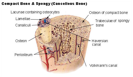
- Anatomy of a Long Bone


Picture of A Long Bone Picture of the compact bone and Spongy
- The main portion of the bone is called the diaphysis.
- Compact bone is highly organized and composed of tubular units called osteons.
- Spongy bone has an unorganized appearance. It contains numerous thin plates separated by unequal spaces.
- Cartilage is not as strong as bone, but it is more flexible because the matrix gel-like and contains many collagenous and elastic fibers.
o Cartilage has no nerves and no blood vessels, making it harder to heal.
o Three types of cartilage:
1. Hyaline cartilage covers the end of the long bone. Is firm and somewhat flexible.
2. Fibro cartilage is stronger than hyaline cartilage because the matrix contains wide rows of thick, collagen fibers.
3. Elastic cartilage is more flexible than hyaline cartilage because the matrix contains mostly elastin fibers.
- Fibrous connective tissue contains rows of cells called fibroblast separated by bundle of collagenous fibers. This tissue makes up ligaments that connect bone to bone and tendons that connect muscles to a bone at joints.

- Bones can grow throughout a lifetime because they are able to respond to stress by changing size, shape, and strength.
- Bones are composed of living tissues
- Cells involved in growth, remodeling, and repair of the bone are:
o Osteblasts-are bone-forming cells. They secrete the organic matrix of bone and promote the deposition of calcium salts into the matrix.
o Osteocytes-are mature bone cells derived from osteoblasts. They maintain structure of bone.
o Osteoclasts-are bone-absorbing cells. They break down bone and assist in depositing calcium and phosphate in the blood.
- Bone Development and Growth
- Ossification refers to the formation of bone.
- Intramembranous ossification: bones develop between sheets of fibrous connective tissue.
- Endochondral ossification: cartilaginous models of the bones are replaced by calcified bone matrix.
- Bone growth is affected by vitamin D, growth hormone, and sex hormones.
- Bone Remodeling and Its Role in Homeostasis
- Bone remodeling is the renewal of bone. Osteoclasts break down bone and osteoblasts re-form bone. Some bone is recycled each year.
- Bone recycling allows the body to regulate blood calcium.
- Bone remodeling also accounts for why bones can respond to stress.
- Bone Repair
- Repair of a fracture required four steps:
o Hematoma formation
o Fibrocartilginous callus
o Bony callus

The Human Skeleton
- Bones of the axial skeleton
- The axial skeleton consists of:
o The skull
o The hyoid bone
o The vertebral column,


The Human Skull Bones of the Face


The Vertebral Column The Rib Cage
- The skull is formed by the cranium, which protects the brain and the facial bones.
- The hyoid bone anchors the tongue and is the site of attachment of muscles involved with swallowing.
- The vertebral column is composed of vertebrae separated by shock-absorbing disks, which make the column flexible. It supports the head and truck, protects the spinal cord, and is a site for muscle attachment.
- The vertebral column consists of 33 vertebrae.
- Rib Cage
- The rib cage is composed of the thoracic vertebrae, ribs, costal cartilages, and sternum. It protects the heart and lungs.
- The rib cage is part of the axial skeleton.
- Protects the heart and lungs
- Bones of the Appendicular Skeleton
- The appendicular skeleton consists of the bones within the pectoral and pelvis girdles and their attached limbs.
- The bodies have left and right pectoral girdles; each consists of a scapula and clavicle.
- The pectoral girdles and upper limbs are adapted for flexibility.
- The bones in the pectoral girdle are:
o Scapula, clavicle
o The upper limb: humerus, radius, ulna, carpals, metacarpals, phalanges
- The pelvic girdle and the lower limbs are adapted for supporting weight; the femur is the longest and strongest bone in the body.
- The bones in the pelvis girdle are:
o Two coxal bones
o The lower limb: Femur, patella, tibia, fibula, tarsals, metatarsals, and phalanges.
- Articulations
- Bones are joined at joints, of which are thee types:
o Fibrous joints-are immovable
o Cartilaginous joints- is slightly movable.
o Synovial joints – are freely movable.
- Movements Permitted by Synovial Joints
- Intact skeletal muscles are attached to bones by tendons that span joints. When a muscle contracts, one bone moves in relation to another bone.
http://academic.kellogg.cc.mi.us/herbrandsonc/bio201_McKinley/f6-4c_gross_anatomy_of__c.jpg
http://www.fluoridealert.org/pesticides/compact.spongy.bone.jpg?4abcc000
http://classes.midlandstech.com/bio112/figure7.5endochondral%20ossification.jpg
http://library.thinkquest.org/J0111100/graphics/skull1.JPG
http://biology.clc.uc.edu/graphics/bio105/skeleton.jpg
http://services.epnet.com/GetImage.aspx/getImage.aspx?ImageIID=4826
http://academic.kellogg.cc.mi.us/herbrandsonc/bio201_McKinley/f7-28_vertebral_column_c.jpg
http://www.medicalook.com/systems_images/Rib_cage.jpg
Chapter 12
Muscular System
- Overview of Muscular System
- Skeletal Muscle Fiber Contraction
- Whole Muscle Contraction
- Muscular Disorders
- Homeostasis
- Picture References
- Overview of Muscular System
- Type Of Muscles

- Humans have three type of Muscles tissues:
o Smooth Muscle- is involuntary, fibers are spindle-shaped cells, each with a single nucleus. Occurs in walls of internal organs.
o Cardiac Muscle- is involuntary, forms the heart wall.
o Skeletal Muscle- is voluntary; fibers are tubular, multinucleated, and striated. Is usually attached by tendons to the skeleton.
- Functions of Skeletal Muscles
o Skeletal muscles support the body.
o Skeletal muscles make bones move.
§ Rigor mortis is the contraction of muscles at death due to ATP.
o Skeletal muscles help maintain a constant body temperature.
o Skeletal muscle contraction assists movement in cardiovascular and lymphatic vessels.
o Skeletal muscles help protect internal organs and stabilize joints.
- Skeletal Muscles of the Body
- Skeletal muscles are well organized.
- Muscles are covered with fascia, a type of connective tissue that extends beyond the muscle and becomes its tendon.
- Tendons quite often extend past a joint before anchoring a muscle to a bone.
- When achieving movement, some muscles are prime movers, some are synergists, and other are antagonists.
- Muscles are named for their size, shape, location, direction of fibers, number of attachments, and action.

The Muscle in the Human Body
- Skeletal Muscle Fiber Contraction
- Muscle Fibers and How They Slide
- Muscle fibers contain myofibrils, and myofibrils contain actin and myosin filaments. Muscle contraction occurs when sarcomeres shorten and actin filaments slide past myosin filaments.
- Nerve impulses travel down motor neurons and stimulate muscle fibers at neuromuscular junctions.
- Thick filaments- is composed of several hundred molecules of the protein myosin.
- Thin filaments- primarily, a thin filament consists of two intertwining strands of the protein actin.
- Sliding filaments- an explanation for muscle contraction based on the movement of actin filaments in relation to myosin filaments.
- Control of Muscle Fiber Contraction
- Muscle fibers are stimulated to contract by motor neurons whose axons are in nerves.
- The role of ATP in muscle contraction is to supplies the energy for muscles contraction.

Skeletal muscle fiber and struture

Neuromuscular Juntion
- Whole Muscle Contraction
- Whole muscle contraction is dependent on muscle fiber contraction.
- Muscles Have Motor Units
- A muscle contains motor units: several fibers under the control of a single motor axon.
- Motor unit contraction is described in terms of a muscle twitch, summation, and tetanus.
- The strength of muscle contraction varies according to recruitment of motor units.
- Summation is increased muscle contraction until maximal sustained contraction is called tetanus.
- A whole muscle typically contains many motor units. As the intensity of nervous stimulation increases, more and more motor units in a muscle are activated. It is known as recruitment.
- “Muscle tone” one desirable effect of exercise. The muscle is firm and solid.
- Energy for Muscle Contraction
- Muscles can use various fuel sources for energy and they have various ways of producing ATP during muscle contraction.
- A muscle fiber has three ways to acquire ATP for muscle contraction
o Creatine phosphate transfers a phosphate to ADP and ATP results.
o Fermentation also produces ATP quickly. Fermentation is associated with an oxygen dept because oxygen is needed to metabolize the lactate that accumulates.
o Cellular respiration provides most of the muscle’s ATP but takes longer because much of the glucose and oxygen must be transported in blood to mitochondria. Occurs during exercise.
- Fast twitch fibers are usually anaerobic and seem to be designed for strength because their motor units contain many fibers.
o Helpful in sport activities like, weight lifting, sprinting or golf clubbing.
o Is anaerobic
o Has explosive power
o Fatigues easily
- Slow twitch fibers have a steadier tug and have more endurance despite more units with smaller number of fibers.
o Helpful with running distances, biking and jogging.
o Is aerobic
o Has steady power
o Has endurance
- Delayed Onset Muscle Soreness
o Appears 24-48 hours after vigorous exercise.
o Many be due to tissue injury that takes several days to heal.
o To prevent “DOMS” try warming up before working out and cooling down completely.
o Avoid making sudden MAJOR changes in a exercise routine.
- Muscular Disorders
- Common Muscular Conditions
o Spasms- are sudden and involuntary muscular contractions most often accompanied by pain.
o Convulsion- multiple spasms of skeletal muscles are seizure.
o Cramps- are strong, painful spasms, especially of the leg and foot, usually due to vigorous activity.
o Facial tics- periodic eye blinking, head turning, or grimacing, are spasms that can be controlled voluntarily, by only with great effort.
o Strains- is caused by stretching or tearing of a muscle
o Sprain- is a twisting of a joint leading to swelling and injury, not only of muscle but also of ligaments.
- Muscular diseases
o Myalgia- refers to achy muscles. Caused by overusing or overstretching of a muscle or group of muscles.
o Fibromyalgia- is a chronic condition whose symptoms include achy pain, tenderness, and stiffness of muscles.
o Other diseases are:
§ Muscular dystrophy
§ Myasthenia gravis

Muscle disorder location in the body
- Homeostasis
- Body Systems Produce Movement
- Movement is essential to maintaining homeostasis.
- The skeletal muscular systems work together to enable body movement.
- The muscles and bones produce movement and protect body parts.
- The bones produce red blood cells and are involved in the regulation of blood calcium levels.
- The muscles produce the heat that gives us a constant body temperature.
http://medicalimages.allrefer.com/large/tissue-types.jpg
http://www.drstandley.com/images/muscular.bmp
http://www.octc.kctcs.edu/gcaplan/anat/images/Image286.gif
http://www.lib.mcg.edu/edu/eshuphysio/program/section2/2ch1/2ch1img/neuromus.jpg
http://www.healiohealth.com/Library/Images/Muscle-Disorders.jpg
Thursday, April 10, 2008
LEECH NEUROPHYSIOLOGY LAB
LEECH NEUROPHYSIOLOGY LAB
· What is the electrode measuring?
Electrodes are what you use to record the activity of the neurons. The generic term "electrode" is defined as a conductor that is used to establish electrical contact with a nonmetallic substance. With the use of florescent dye the neurons can be seen using a dissection microscope.
· Why use leeches in neurophysiology experiments?
Leeches have been useful in many experiments in the function of the nervous system. Leeches have a simple nervous system so it’s easier to understand. Many first discoveries were found in leeches. Leeches has 21 segmental ganglia, each containing 175 pairs of neurons. The reasonably small number and the large size of the neurons have made leeches favorite subjects of neurobiologists.
· What is the difference between a sensory and a motor neuron?
Sensory neurons take nerve impulses or messages from a sensory receptor to the CNS. The Motor neuron takes nerve impulse away from the CNS to effectors. The difference between both is that they both take nerve impulses to different locations. The sensory sends messages to the CNS and motor neuron takes it away from the CNS.
· Do you think a leech experiences pain? What is pain?
Leeches have a nervous system so I would believe that they feel some sort of pain during experiences. During this experience, the leech was dipped in 20% ethanol solution. It can be use for smaller creature as an anesthesia like a leech that breathes through their skin. Pain is an unlikable sensation. When there is harm done to our bodies to any creature’s bodies it can cause a discomfort or agony.
· What were the two most interesting things about doing this lab?
To dissects a leech online. After doing this experiment I never knew anything about leeches and how useful they have been in discovering many functions with the nervous system. To learn more about neurons and how it looked after I put the florescent dye in and then the light and how each neuron look different from one another. The pictures of the neurons were specific because I felt like I was really doing the experiment even if it was on a computer.
· Anything you found confusing or didn't like about the lab?
It took me awhile to figure out how to actually get a neuron at first but other than that it was a great lab!

an Image of the leech with the oscillope trace.

Cell Types that are inside a Leech's Nervous System


Wednesday, April 9, 2008
Chapter 13 &14
Chapter 13
Nervous System
Table of Content
- Overview of the Nervous system
- The central nervous system
- The limbic system and higher mental functions
- The peripheral nervous system
- Drug abuse
- Picture references


- Overview of the Nervous system
- Has two major divisions: the central nervous system and the peripheral nervous system.
- The nervous system has three major functions:
- Reception of input
- Integration of data
- Generate motor output
- Nervous Tissue
- Two types of cells: neurons and neuroglia.
- Neurons-transmit nerve impulse between parts of the nervous system.
- Neuroglia- support and nourish neurons.
- Neuron structure
- The type of neurons
- Sensory neurons-are special structures that detect changes in the environment.
- Interneuron-lies entirely within the CNS.
- Motor neurons-takes nerve impulses away from the CNS to an effectors.
- Effectors carry out our responses to environmental changes, whether they are external or internal.
- Composed of dendrites, a cell body, and axon.

Myelin Sheath
- Myelin Sheath
- Long axons are covered by a myelin sheath.
- The Nerve impulse
Resting potential-more Na+ outside the axon and more K+ inside the axon. The axon does not conduct an impulse.
Action potential- a change in polarity across the axonal membrane as a nerve impulse occurs.
- The synapse
- When a neurotransmitter is released into a synaptic cleft, transmission of a nerve impulse occurs.
- Neurotransmitter transmits from on neuron to the next so neurons don’t physically touch.

Synapse
- The central nervous system

CNS
- The spinal cord and the brain make up the CNS.
- Where sensory information is received and motor control is initiated.
- Both the brain and spinal cord are protected in a membrane known as meinges.
- The Spinal Cord
- Extends from the base of the brain through a large opening in the skull called the foramen magnum and into the vertebral canal formed by openings in the vertebrae.
- Grey matter of the spinal cord contains neuron cell bodies.
- White matter consists of myelinated axons that occur in tracts.
- The spinal cord provides a means of communication between the brain and the peripheral nerves that leave the cord.
- Conduction to and from brain; carries out reflex actions.

Spinal Cord
- The Brain
- The cerebrum is the largest portion of the brain in humans. Is the last center to receive sensory input and carry out integration before commanding voluntary motor responses.
- Four major parts of the brain are: cerebrum, diencephalon, cerebellum, and the brain stem.
- Has two cerebral hemispheres connected by the corpus callosum.
- Shallow grooves called sulci divide each hemisphere into lobes
- Frontal lobe-the most ventral of the lobes.
- Parietal lobe- is dorsal to the frontal lobe.
- Occipital lobe- is dorsal to the parietal lobe.
- Temporal lobe- lies inferior to the frontal and parietal lobe.
- Each lobe is associated with particular functions.
- The cerebral cortex- is a thin but highly convoluted outer layer of gray matter that covers the cerebral hemispheres.
- Covers the cerebrum.
- Diencephalon- contains the hypothalamus and thalamus, maintains homeostasis, and receives sensory input.
- Cerebellum- sends out motor impulses by way of the brain stem to the skeletal muscles, produces smooth, coordinated voluntary movements.
- Brain stem- contains the midbrain, pons, and medulla oblongata, relay station and medulla had reflex centers.

the Lobes in the Brain
- The limbic system and higher mental functions
- Limbic system
- Functions of limbic system-
- Blend primitive emotions and higher mental functions into a united whole.
- The amygdala can cause experiences to have emotional overtones, and it creates the sensation of fear.
- The hippocampus plays a crucial role in learning and memory. It’s involved in storing and retrieving memories.

Limbic System
- Higher mental Functions
- Memory is the ability to hold a thought in mind or to recall events from the past, ranging from a word we learned only yesterday to an early emotional experience that has shaped our lives.
- Learning takes place when we retain and utilize past memories.
- Type of memory:
- Short term memory
- Long term memory
- Semantic memory
- Episodic memory
- Skill memory
- Long-term memory is stored in bits and pieces throughout the sensory association areas of the cerebral cortex.
- Language depends on semantic memory.

4. The peripheral nervous system
- The PNS contains only nerves and ganglia.
- Cranial nerves take impulses to and from the brain. There are 12 pairs of cranial nerves.

Cranial Nerves
Spinal nerves take impulses to and from the spinal cord. There are 31 pairs of spinal nerves.
- The PNS is divided into the somatic system and the autonomic system.
- Somatic System
- Serve the skin, skeletal muscle, and tendons.
- Reflexes are automatic responses to a stimulus in the somatic system. A reflex occurs quickly, without us thinking!!
- Autonomic System
- Regulates the activity of cardiac and smooth muscles and glands.
- A function involuntarily, has two systems that normally cause opposite responses.
- Sympathetic Division
- Parasympathetic division
- Drug abuse
- Drug abuse is apparent when a person takes a drug at a dose level and under circumstances that increase the potential for a harmful effect.
- Drug abuse affect the limbic system
- And promote or decrease the action of a particular neurotransmitter.
- Alcohol
- Is dangerous to the body and brain.
- CNS, alcohol acts as a depressant and influences many brain regions and neurotransmitter.
- Nicotine
- Acts as a stimulant.
- In the PNS, nicotine imitations acetylcholine and increases skeletal muscles activity, heart rate and blood pressure.
- Cocaine
- Powerful stimulant in the CNS.
- Methamphetamine
- Acts as a stimulant.
- Death can occur.
- Heroin
- Acts as a depressant.
- Is the most abuse opiate because it is rapidly delivered to the brain.
- Marijuana
- Acts as a psychoactive.
- Has influence in the brain.
- Has a reaction to the CNS.
- Has some medical use to people who suffer from illness.
Picture References:
http://www.issaonline.com/trial/unit1/images/nervoussystem.jpg
http://z.about.com/f/p/440/graphics/images/en/8679.jpg
http://academic.kellogg.cc.mi.us/herbrandsonc/bio201_McKinley/f14-3a_structures_in_a__c.jpg
http://medicalimages.allrefer.com/large/central-nervous-system-1.jpg
http://people.eku.edu/ritchisong/spinalcord5.gif
http://www.wright.edu/academics/honors/institute/images/brain_diagram.jpg
http://www.macalester.edu/psychology/whathap/ubnrp/dopahypoweb04/limbicsystem.jpg
http://mybrainic.com/images/otak_1_eng.gif
http://www.macalester.edu/psychology/whathap/UBNRP/vagus05/Vagus_pics/cranialnerves.jpg
Chapter 14
Senses
Table of Content
- Sensory Receptors and Sensations
- Proprioceptors and Cutaneous Receptors
- Senses of Taste and Smell
- Sense of Vision
- Sense of Hearing
- Sense of Equilibrium
- Picture References
- Sensory Receptors and Sensations
· Function of a sensory receptor is to detect certain type of stimuli.
· Type of Sensory Receptors
o Chemoreceptor-respond to chemical substances in the immediate vicinity. Ex. Receptors for taste and smell.
o Photoreceptor-respond to light energy. Ex. Vision. Eyes.
o Mechanoreceptor- are stimulated by mechanical forces, which most often result in pressure of some sort. Ex. From pressure, sound waves and gravity. Hearing.
o Thermoreceptor- sensory receptor that is sensitive to changes in temperature. Ex. Body temperature.
· How Sensation Occurs
o Sensation is the conscious perception of stimuli that occurs after sensory receptors generate a nerve impulse that arrives at the cerebral cortex.
- Proprioceptors and Cutaneous Receptors
· Proprioceptors
o Are mechanoreceptors involved in reflex actions that maintain muscle tone and thereby the body’s equilibrium and posture.
o To assist the brain in knowing the position of the limbs and space
· Cutaneous Receptors
o Are found in the skin
o Are for touch, pressure, temperature, and pain.
· Pain Receptors

Sensory receptors in human skin
- Senses of Taste and Smell
· Taste and smell are called chemical senses because their receptors are sensitive to molecules in the food we eat and the air we breathe.
· Taste and smell are due to chemoreceptors that are stimulated by molecules in the environment.
· Sense of Taste
o About 3,000 taste buds are located mostly on the tongue.
o Most taste buds lie along the walls of the papillae.
o At least four primary types of taste:
o Sweet
o Sour
o Salty
o Bitter
o Umami, the fifth taste, may exist for certain flavor.


Type of taste locations Sense of Taste and their cells
o Taste buds open at a taste pore.
o Microvilli of taste cells have receptor proteins fro molecules that cause the brain to distinguish, sweet, sour, salty, and bitter tastes.
· Sense of Smell
o Approximately 80%-90% of what we perceive as “taste” actually is due to sense of smell
o The cilia of olfactory cells have receptor proteins for molecules that cause the brain to distinguish odors.
o Olfactory cells are located high in the roof of the nasal cavity.
o The olfactory bulbs have direct connections with the limbic system and its center for emotions and memory.


A Human Eye
· Vision depends on the eye, the optic nerves, and the visual areas of the cerebral cortex.
· Anatomy and Physiology of the Eye
o The eye has three layers, or coats:
o The sclera (outer layer) protects and supports the eyeball.
o The choriod (middle pigmented layer) absorbs stray light rays.
o The retina (inner layer) contains the rod cells (sensory receptors for dim light) and cone cells (sensory receptors for bright light and color).
o The lens brings the light rays to focus on the retina. To see a close object, visual accommodation occurs as the lens rounds up.
o The pathway for vision begins once light has been focused on the photoreceptors in the retina.
o Functions of photoreceptors
o Rod cells
o Cone cells
§ Both have an outer segments joined to an inner segment by a stalk.
· Abnormalities of the Eye
o Color blindness
o Misshapen eyeballs
o Complete color blindness is extremely rare.
o Nearsighted people can see close objects better than they can see objects at a distance.
o Farsighted people can see distant objects better than they can see close objects.
o Astigmatism, is when the cornea or lens is uneven, the image is fuzzy. The light rays cannot be evenly focused on the retina.

The Human Ear
· The ear has two sensory functions:
o Hearing and balance.
o Both located in the inner ear.
o Each consists of hair cells with stereocilia that are sensitive to mechanical.
o There are mechanoreceptors.
o Hearing depends on the ear, the cochlear nerve, and the auditory areas of the cerebral cortex.
· Anatomy and Physiology of the Ear
o The ear has three parts:
o In the outer ear, the pinna and the auditory canal direct sound waves to the middle ear.
o In the middle ear, the tympanic membrane and the ossicles (malleus, incus, and stapes) amplify sounds waves.
o In the inner ear, the semicircular canals detect rotational equilibrium; the utricle and saccule detect gravitational equilibrium; and the cochlea house the spiral organ, which contains mechanoreceptors for hearing.
o The auditory pathway begins when the outer ear receives and the middle ear amplifies sound waves that then strike the oval window membrane.
o Mechanoreceptors allow us to hear and are located in the inner ear. They function by sensitive to mechanical stimulation.
- Sense of Equilibrium
· The ear also contains mechanoreceptors for equilibrium.
· Rotational Equilibrium Pathway
· Mechanoreceptors in the semicircular canals detect rotational and/or angular movement of the head. (Rotational equilibrium).
· Gravitational Equilibrium Pathway
· The receptors are located: Utricle and saccule
· It’s function is: Utricle and saccule contain hair cells with sterocilia embedded in an otolithic membrane; when the head bends, otoliths are displaced causing the sterocilia to bend.
Picture References:
http://fig.cox.miami.edu/~cmallery/150/neuro/c7.49.3.skin.jpg
http://library.thinkquest.org/3750/images/tastebud.gif
http://classes.midlandstech.com/bio112/taste%20buds.jpg
Subscribe to:
Posts (Atom)
